Cervical spondylosis may compress the nerves exiting your neck before warning signs become apparent. As the cushioning discs between cervical vertebrae lose hydration and height, bone spurs can form along vertebral edges and narrow the spaces where spinal nerves exit, potentially producing persistent neck pain, stiffness, and radiating arm symptoms that prompt many patients to seek medical evaluation.
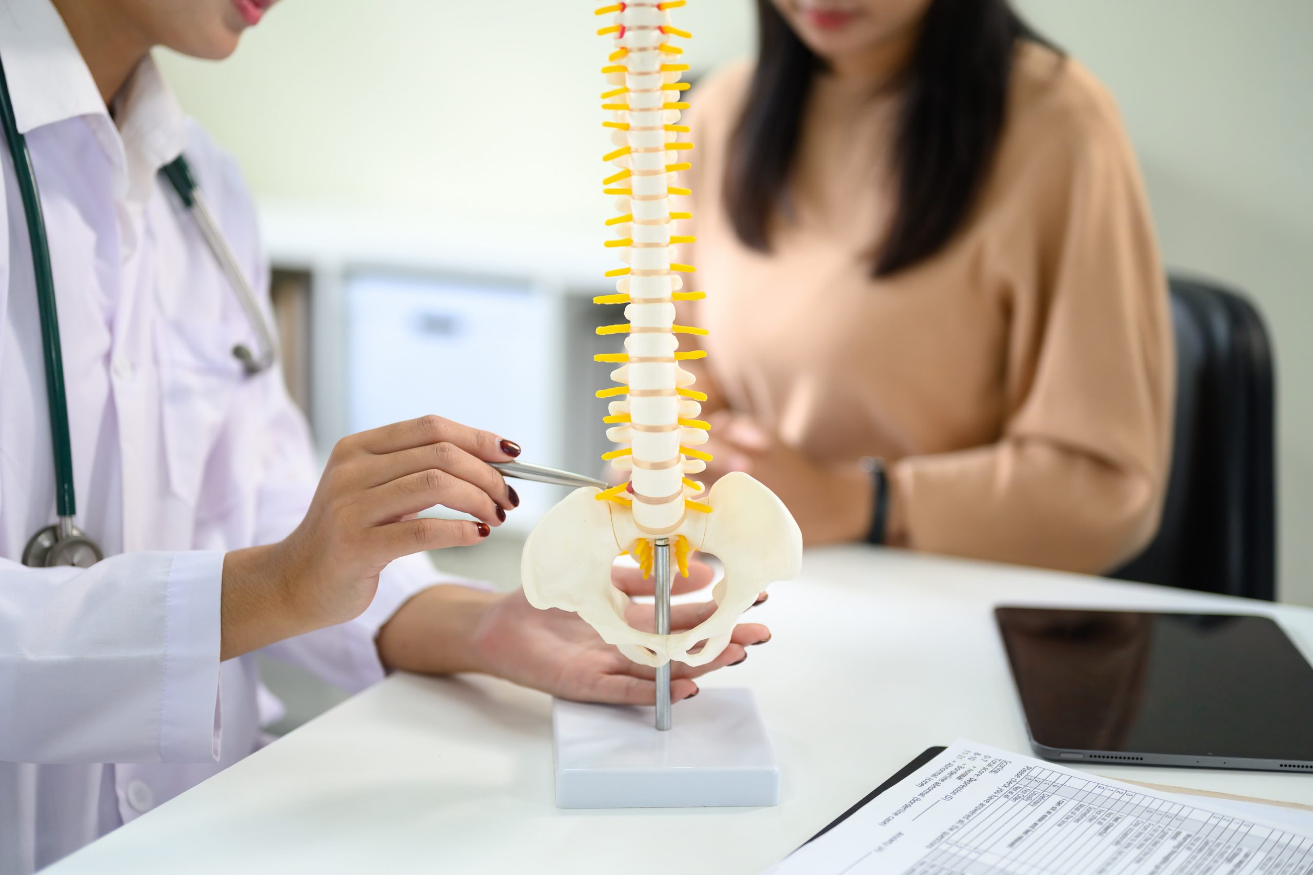
The cervical spine comprises seven vertebrae (C1–C7) stacked vertically, separated by intervertebral discs that absorb shock and allow movement. Each vertebra has openings called foramina through which nerve roots pass to supply sensation and motor function to your shoulders, arms, and hands. When cervical spondylosis narrows these passages or compresses the spinal cord itself, symptoms can extend beyond simple neck discomfort into neurological territory requiring thorough assessment.
How Cervical Spondylosis Develops
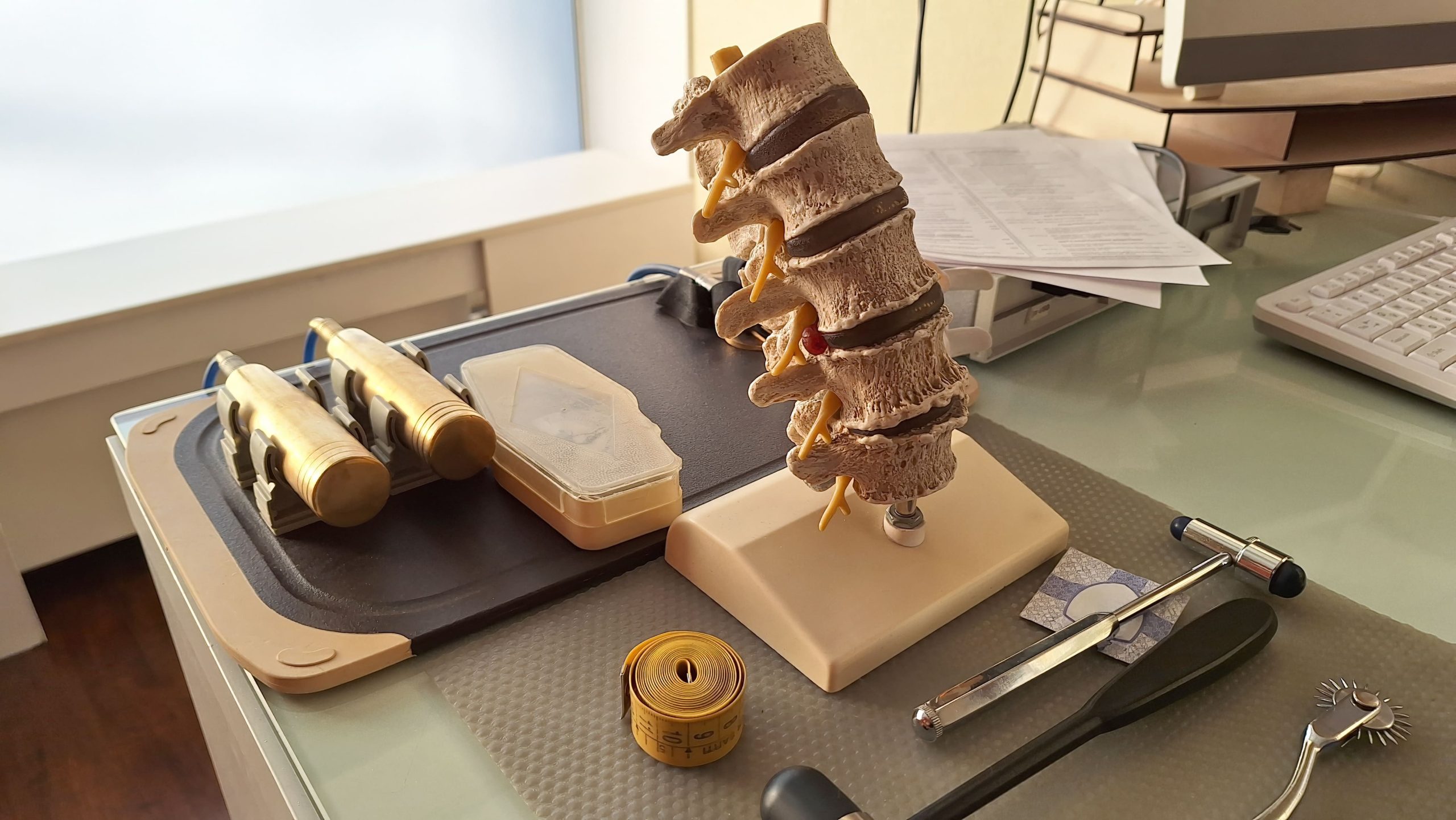
Intervertebral discs contain a gel-like nucleus surrounded by tough fibrous rings. Over time, the nucleus often loses water content, reducing disc height and its ability to cushion vertebrae during movement. This height loss can increase stress on facet joints—the small joints connecting adjacent vertebrae at the back of the spine, potentially accelerating cartilage breakdown and bone spur formation.
Bone spurs, or osteophytes, represent the body’s attempt to stabilise a spine experiencing increased mechanical stress. Whilst this response is protective, osteophytes can protrude into the spinal canal or neural foramina. The ligamentum flavum, a tissue connecting vertebral arches, may also thicken with age, further reducing available space for neural structures.
Occupational factors can influence progression rates. Prolonged desk work, frequent overhead lifting, and jobs requiring repetitive neck movements may accelerate degenerative changes. Previous neck injuries, even minor whiplash events occurring years earlier, may create vulnerable segments that are more susceptible to spondylotic changes.
Recognising the Symptoms
Localised Neck Symptoms
Neck stiffness typically worsens after prolonged positioning—morning stiffness after sleep or end-of-day tightness following desk work. Pain often localises to the lower cervical region (C5–C7), where movement demands are highest. Grinding or popping sensations during neck rotation may reflect bone-on-bone contact or osteophyte movement.
Headaches originating from cervical spondylosis typically start at the base of the skull and radiate forward. These cervicogenic headaches differ from migraines; while both can be one-sided, cervicogenic headaches typically do not cause nausea, vomiting, or sensitivity to sound (phonophobia), and they usually worsen with sustained neck positions rather than light or sound exposure.
Radicular Symptoms
When nerve roots become compressed, symptoms generally follow specific patterns based on which nerve is affected:
- C5 nerve root: Shoulder pain with deltoid weakness; difficulty raising the arm sideways
- C6 nerve root: Pain radiating to the thumb and index finger; weakened wrist extension and biceps
- C7 nerve root: Pain extending to the middle finger; triceps weakness affecting pushing movements
- C8 nerve root: Pain in the ring and small fingers; grip weakness and difficulty with fine motor tasks
Numbness, tingling, or burning sensations in these distributions suggest nerve irritation rather than simple muscular neck pain.
Myelopathic Symptoms
Cervical myelopathy occurs when the spinal cord itself becomes compressed—a more serious presentation requiring prompt evaluation. Warning signs include:
- Difficulty with balance or coordination when walking
- Clumsiness in hands, particularly with buttons or writing
- Electric shock sensations running down the spine with neck flexion (Lhermitte’s sign)
- Leg stiffness or weakness
- Bladder urgency or hesitancy
These symptoms may indicate spinal cord dysfunction and warrant urgent medical assessment.
Diagnostic Approaches
Clinical Examination
Physical examination assesses neck range of motion, identifies painful movements, and maps neurological function. Spurling’s test—applying downward pressure on the head whilst tilting it toward the affected side—may reproduce radicular pain when nerve root compression exists. Hoffman’s sign, hyperactive reflexes, and gait abnormalities can suggest myelopathy.
Muscle strength testing grades power from 0 (no movement) to 5 (normal strength) across specific muscle groups, helping localise which nerve roots may be affected. Sensory testing with light touch and pinprick maps dermatomal distributions.
Imaging Studies
X-rays can reveal disc space narrowing, osteophyte formation, and alignment abnormalities. Flexion-extension views assess spinal stability during movement.
MRI provides detailed visualisation of soft tissues—intervertebral discs, spinal cord, and nerve roots. MRI can demonstrate disc herniations, cord compression, and signal changes within the spinal cord suggesting damage. This modality is particularly valuable for surgical planning.
CT scans offer detailed bone imaging, useful for evaluating osteophyte size and location. CT myelography, involving contrast injection into the spinal fluid space, provides good visualisation when MRI cannot be performed.
Nerve conduction studies and electromyography (EMG) help assess electrical function of nerves and muscles, aiding in differentiating cervical radiculopathy from peripheral nerve entrapments like carpal tunnel syndrome.
💡 Did You Know?
The cervical spine supports a head weighing approximately 4.5–5 kg when properly aligned. Forward head posture — common with prolonged smartphone or computer use, places additional mechanical stress on cervical structures and may accelerate degenerative changes over time.
Non-Surgical Treatment Options
Physical Therapy
Structured physiotherapy programmes address muscle imbalances contributing to symptoms. Strengthening deep neck flexors aims to improve cervical stability, whilst stretching tight posterior neck muscles and pectorals may help correct forward head posture. Manual therapy techniques including mobilisation and manipulation may improve joint mobility in appropriate candidates.
Postural retraining focuses on maintaining neutral cervical alignment during daily activities. Ergonomic modifications—monitor height adjustment, keyboard positioning, and regular movement breaks—aim to reduce sustained mechanical stress.
Medications
Anti-inflammatory medications are commonly used to reduce pain and inflammation during acute flares. Long-term use requires monitoring for gastrointestinal and cardiovascular effects.
Neuropathic pain medications including gabapentin and pregabalin may help manage nerve-related symptoms by addressing abnormal nerve signalling.
Muscle relaxants can address associated muscle spasm but may cause sedation, often limiting daytime use.
Interventional Procedures
Cervical epidural steroid injections deliver anti-inflammatory medication directly around compressed nerve roots. Performed under fluoroscopic guidance, these injections aim to reduce inflammation and pain, potentially allowing more effective participation in rehabilitation.
Facet joint injections target pain originating from degenerated facet joints rather than nerve compression.
Radiofrequency ablation uses heat to interrupt pain signals from facet joints, which may provide extended relief for selected suitable patients.
Surgical Considerations
When Surgery Becomes Necessary
Surgery is typically considered for:
- Progressive neurological deficits despite conservative treatment
- Significant myelopathy with functional impairment
- Severe radiculopathy unresponsive to non-surgical measures over several months
- Spinal instability
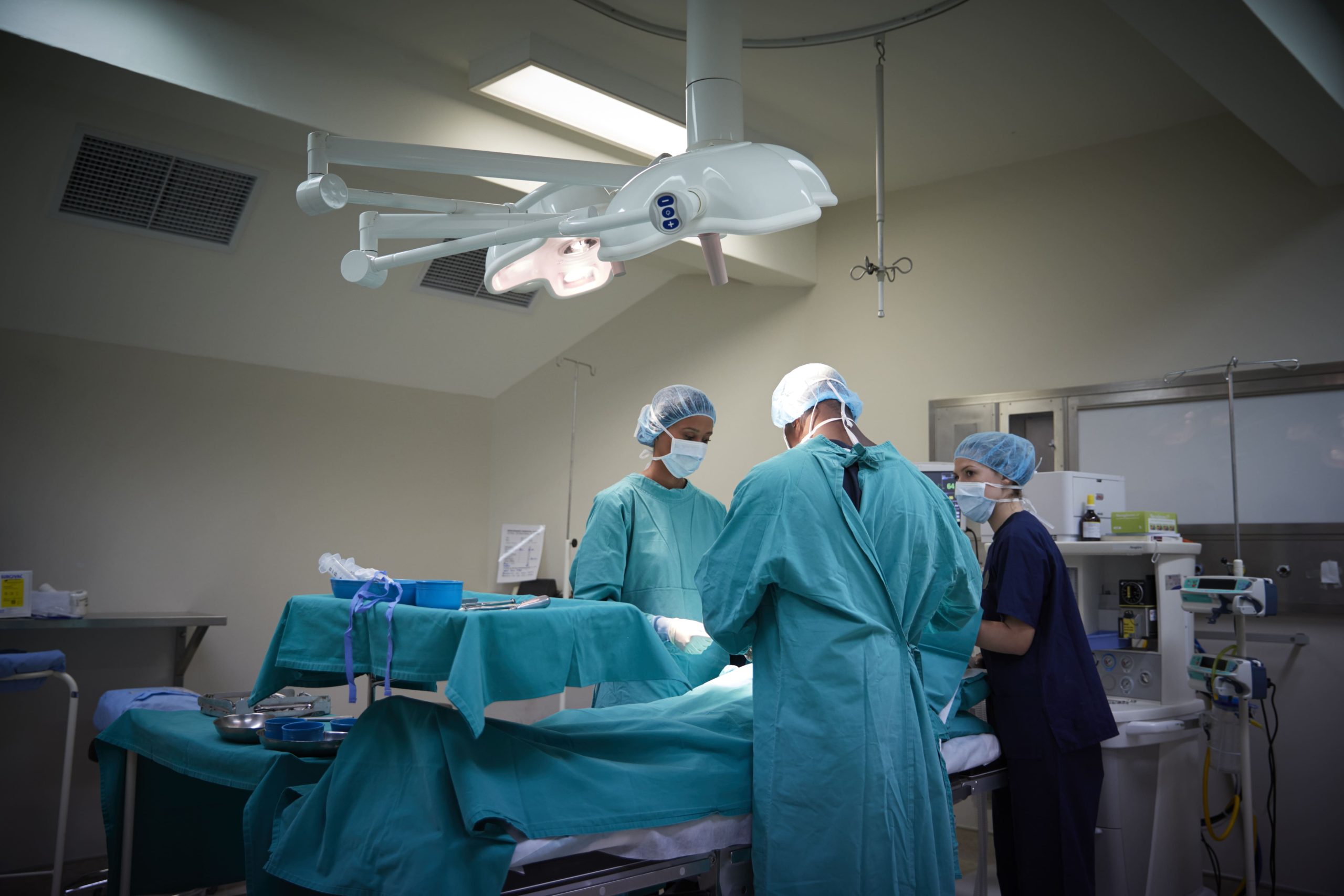
Surgical Approaches
Anterior cervical discectomy and fusion (ACDF) involves approaching the spine through a small incision in the front of the neck. The damaged disc is removed, nerve roots are decompressed, and a bone graft or cage is inserted with the goal of fusing adjacent vertebrae. Metal plates and screws aim to provide immediate stability.
Cervical disc replacement substitutes an artificial disc for the damaged one, designed to help preserve motion at the treated level. This option may suit patients with single-level disease and healthy adjacent segments.
Posterior cervical laminectomy removes the lamina (back portion of vertebrae) to create more space for the spinal cord. This approach is often considered for multilevel disease with cord compression.
Posterior cervical laminoplasty hinges the lamina open rather than removing it completely, aiming to maintain spinal stability whilst decompressing the cord.
Combined anterior-posterior surgery is sometimes utilised to address severe multilevel disease requiring both decompression and stabilisation.
⚠️ Important Note
Myelopathic symptoms including progressive walking difficulty, hand clumsiness, or bladder dysfunction warrant urgent specialist evaluation. Delayed treatment of significant cord compression may lead to permanent neurological deficits.
Daily Strategies for Symptom Management
Modify workstation setup by positioning your monitor at eye level with the screen approximately an arm’s length away. Use a document holder beside the monitor rather than looking down at papers.
Apply heat or ice strategically—heat may help relax tight muscles and support blood flow for chronic stiffness, whilst ice can help reduce inflammation during acute flares.
Practise chin tucks by drawing your chin straight back whilst keeping your eyes level, holding for several seconds. This exercise is designed to strengthen deep neck flexors and help counteract forward head posture.
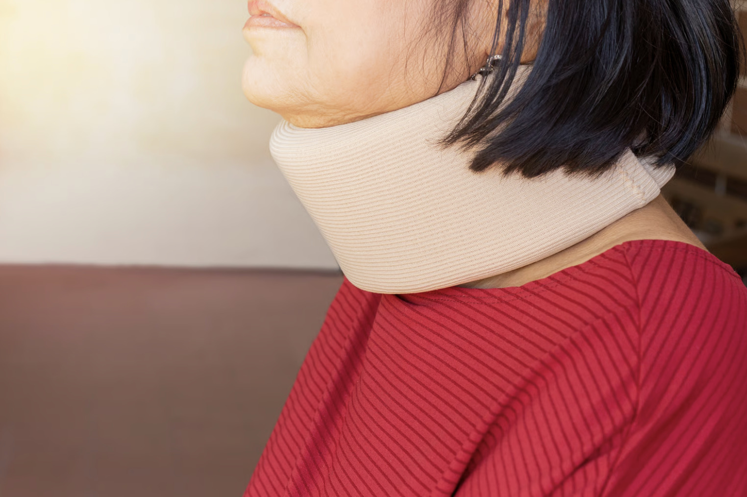
Sleep with appropriate support using a cervical pillow that helps maintain neutral spine alignment. Avoid stomach sleeping, which forces prolonged neck rotation.
Take movement breaks every 30–45 minutes during desk work, performing gentle neck range of motion exercises to help reduce sustained loading.
When to Seek Professional Help
- Neck pain persisting beyond several weeks despite rest and over-the-counter medications
- Numbness, tingling, or weakness radiating into your arm or hand
- Pain that wakes you from sleep or prevents comfortable positioning
- Difficulty gripping objects or noticing increased hand clumsiness
- Changes in walking pattern or balance
- Bladder or bowel function changes
- Symptoms following trauma, even minor incidents
Commonly Asked Questions
Can cervical spondylosis be reversed?
Structural changes including disc degeneration and bone spur formation cannot be reversed. However, symptoms can often be managed, and progression may be slowed through appropriate treatment, postural modification, and regular exercise. Many patients maintain good function with conservative measures alone.
How do I know if my neck pain needs surgery?
Surgery is typically considered when conservative treatments fail to control symptoms adequately, when progressive neurological deficits develop, or when imaging demonstrates significant spinal cord compression with corresponding clinical findings. Many patients with cervical spondylosis respond well to non-surgical management.
Will my symptoms get worse over time?
Cervical spondylosis is a progressive condition, but the rate varies considerably between individuals. Active management including regular exercise, ergonomic modifications, and maintaining a healthy weight may help slow progression. Some patients experience stable symptoms for years, whilst others may require escalating treatment.
Is it safe to exercise with cervical spondylosis?
Appropriate exercise is generally beneficial and recommended. Low-impact activities like swimming, walking, and supervised strengthening programmes support cervical spine health. Avoid high-impact activities, heavy overhead lifting, and exercises that require extreme neck positions. A physiotherapist can design a safe programme suited to your specific condition.
Can poor posture alone cause these symptoms?
Poor posture can accelerate degenerative changes and may worsen symptoms from existing cervical spondylosis. However, spondylosis involves structural changes beyond simple postural effects. Addressing posture is an important part of management but does not eliminate underlying degenerative pathology.
Next Steps
Many cases of cervical spondylosis are managed without surgery, but radiating arm pain, numbness, hand weakness, or changes in balance require proper evaluation to help rule out nerve root or spinal cord compression. Clinical examination combined with MRI or CT imaging helps identify the potential source of symptoms and determine whether conservative management, interventional procedures, or surgical decompression is most appropriate.
If you are experiencing persistent neck pain with radiating arm symptoms, numbness, hand weakness, or difficulty with balance, consult our Orthopaedic Surgeon or Neurosurgeon for a comprehensive evaluation.