Spondylolisthesis: Guide to Diagnosis and Treatment in Singapore

Living with lower back pain that radiates down your legs can be frustrating and debilitating. If you’ve been experiencing persistent discomfort affecting your daily activities, you may be dealing with a condition where one vertebra (the individual bones making up your spine) slips forward over the bone below it.

This condition affects people of all ages, from young athletes to older adults. Understanding your condition is the first step towards management. Some patients with spondylolisthesis can achieve relief and return to activities with appropriate treatment.

This guide provides information about spondylolisthesis, including its causes, symptoms, diagnostic methods, and treatment options available.

Dr Ng Zhi Xu
Senior Consultant Neurosurgeon
Dr Roy Koh Kiok Miang
Senior Consultant Neurosurgeon
Dr Victor Wang Tzong-Jing
Senior Consultant Orthopaedic Surgeon, Spine & Orthopaedic Surgery

What is Spondylolisthesis?

Spondylolisthesis is a spinal condition characterised by the forward displacement of one vertebra over the vertebra directly beneath it. The term comes from Greek words meaning “spine” and “slipping.” This slippage commonly occurs in the lower back, particularly at the L4-L5 or L5-S1 vertebral levels (specific bones in your lower spine).

When a vertebra slips out of position, it can narrow the spinal canal (the protective channel housing your spinal cord) and compress nearby nerve roots (the nerves branching from your spinal cord). This compression may lead to pain, numbness, or weakness in the lower back and legs. Symptom severity can vary with the degree of slippage.

Spondylolisthesis is seen amongst the adult population, particularly amongst individuals who engage in activities requiring repetitive hyperextension of the spine (bending backwards repeatedly), such as gymnastics, diving, and weightlifting. Certain populations may have genetic predispositions to developing this condition.

Types of Spondylolisthesis

Isthmic Spondylolisthesis

This type is caused by a defect or fracture in the pars interarticularis (a small segment of bone connecting the facet joints—the joints that link adjacent vertebrae together). This defect, called spondylolysis, can occur due to repetitive stress or trauma. When bilateral pars defects are present (defects on both sides of the vertebra), the vertebra becomes unstable and may slip forward. Isthmic spondylolisthesis may affect adolescents and young adults involved in sports.

Degenerative Spondylolisthesis

Age-related wear and tear on the spinal discs and facet joints can lead to this type. As discs lose height and facet joints develop arthritis, the spine becomes less stable. This form may affect older adults. The L4-L5 level (the joint between the fourth and fifth lumbar vertebrae in the lower back) is often involved.

Congenital Spondylolisthesis

This type is present from birth and results from abnormal bone formation during foetal development. Malformed facet joints may not provide sufficient stability, allowing vertebral slippage. Symptoms may not appear until later childhood or adolescence.

Traumatic Spondylolisthesis

A sudden injury or fracture to the vertebrae can cause acute slippage. High-impact trauma (such as motor vehicle accidents or significant falls) causes this type. Immediate medical attention may be necessary.

Pathological Spondylolisthesis

Underlying diseases that weaken bone structure can lead to this type. Conditions such as osteoporosis (a condition making bones less dense and more prone to fracture), tumours, or infections may compromise vertebral integrity and stability.

Causes

  • Pars interarticularis defect: A crack or stress fracture in this small bone segment (a thin piece of bone connecting the upper and lower joints of each vertebra) can allow one vertebra to slip forward over another.
  • Disc degeneration: Age-related breakdown of intervertebral discs (the cushion-like pads between your vertebrae) can reduce spinal stability.
  • Facet joint arthritis: Worn facet joints (the small joints connecting each vertebra and helping control movement) may not provide appropriate support for vertebral alignment.
  • Congenital vertebral abnormalities: Structural defects present from birth predispose to slippage.
  • Bone-weakening conditions: Diseases affecting bone density, such as osteoporosis or bone tumours, compromise spinal integrity.

Risk Factors

  • Age: Degenerative spondylolisthesis may increase after middle age.
  • Gender: Women may experience degenerative spondylolisthesis, possibly due to hormonal factors affecting joint laxity (looseness or flexibility of the joints).
  • Athletic activities: Sports requiring repetitive lumbar hyperextension (excessive backward bending of the lower back), such as gymnastics, cricket fast bowling, and weightlifting, may increase risk.
  • Family history: Genetic predisposition plays a role in some types.
  • Occupation: Jobs involving heavy lifting, prolonged standing, or repetitive bending stress the spine.
  • Obesity: Excess weight places stress on the lumbar vertebrae.
  • Previous spinal surgery: Prior procedures may alter spinal biomechanics (the way your spine moves and supports your body).

Signs and Symptoms

Spondylolisthesis symptoms vary considerably depending on the degree of vertebral slippage and nerve involvement. Some individuals with mild slippage experience no symptoms.

  • Intermittent lower back pain worsening with activity
  • Muscle tightness or stiffness in the lower back and hamstrings
  • Mild discomfort after prolonged standing or walking
  • Slight decrease in flexibility when bending forward or backward
  • Occasional aching sensation in the buttocks


Mild symptoms may respond to conservative measures and may not significantly impact daily activities. Some patients at this stage can manage their condition with lifestyle modifications and exercise.

  • Persistent lower back pain affecting daily activities
  • Pain radiating into the buttocks and thighs
  • Numbness or tingling sensations in the legs
  • Muscle weakness in the lower extremities
  • Difficulty maintaining posture
  • Increased pain when standing upright or walking (neurogenic claudication—pain caused by nerve compression worsening with activity)
  • Relief when bending forward or sitting


At this stage, symptoms may begin to interfere with work, exercise, and recreational activities. Relief experienced when leaning forward may occur because this position opens up the spinal canal, reducing pressure on the nerves.

  • Constant pain disrupting sleep
  • Significant nerve pressure causing pronounced leg weakness
  • Difficulty walking or standing for even short periods
  • Loss of bladder or bowel control (cauda equina syndrome—requires emergency care)
  • Visible changes in posture or gait
  • Muscle wasting in the legs


Severe symptoms may indicate significant nerve compression and may require more intensive treatment. Cauda equina syndrome is a medical emergency requiring immediate surgical intervention.

When to See a Doctor

Understanding when to seek medical attention for back pain can be challenging. Consider consulting an orthopaedic surgeon or neurosurgeon if you experience specific symptoms.

Symptoms Requiring Prompt Attention

  • Back pain accompanied by leg weakness or numbness
  • Progressive symptoms despite rest and home remedies
  • Pain that wakes you from sleep
  • Difficulty walking or changes in your gait (the way you walk)
  • Loss of bladder or bowel control (seek emergency care immediately)
  • Pain following significant trauma or injury

Timing for Consultation

Early diagnosis can improve outcomes. If your lower back pain persists for more than a few weeks despite rest and over-the-counter medications, schedule an appointment. Athletes experiencing recurrent back pain during training should seek evaluation promptly. Qualified healthcare professionals can assess your specific situation and determine the course of action tailored to your individual symptoms and risk factors.

What to Expect at Your First Consultation

  • Your orthopaedic surgeon or neurosurgeon will conduct a thorough assessment, including detailed medical history, physical examination, and neurological testing (checks to evaluate nerve function, such as testing reflexes, muscle strength, and sensation).
  • Be prepared to discuss when symptoms began, what aggravates or relieves your pain, and how symptoms affect your daily life.
  • Bring any previous imaging studies (such as X-rays or MRIs) or medical records.

Experiencing these symptoms?


Schedule a consultation with an orthopaedic surgeon or neurosurgeon to receive an accurate diagnosis and a personalised treatment plan.

Diagnosis and Testing Methods

Accurate diagnosis of spondylolisthesis involves a combination of clinical evaluation and imaging studies (such as X-rays, MRI scans, and CT scans).

Physical Examination

Your orthopaedic surgeon or neurosurgeon will assess your posture, range of motion, and gait. Specific tests evaluate nerve function, muscle strength, and reflexes. Palpation of the spine may reveal a step-off deformity at the level of slippage.

X-rays

Plain radiographs remain a diagnostic tool. Standing X-rays of the lumbar spine, including flexion and extension views, can reveal vertebral alignment and the degree of slippage. The Meyerding classification system grades spondylolisthesis from Grade I (minor slippage) to Grade V (complete slippage).

Magnetic Resonance Imaging (MRI)

MRI (a scan using magnets to create detailed images) provides detailed images of soft tissues, including intervertebral discs, spinal cord, and nerve roots. This imaging can be valuable for evaluating nerve compression and planning treatment. MRI does not involve radiation exposure.

Computed Tomography (CT) Scan

CT scanning (a detailed X-ray that produces cross-sectional images) provides detailed visualisation of bone structures and can be helpful in assessing pars interarticularis defects (small fractures in the spine). CT may be combined with myelography (an X-ray with contrast dye) for enhanced nerve root visualisation in complex cases.

Bone Scan

Nuclear bone scanning (a test using a small amount of radioactive material) detects areas of increased bone activity and can help determine if a pars defect is acute (healing potential) or chronic. This information can influence treatment decisions, particularly in young patients. Imaging results can arrive within several days to one week. Your orthopaedic surgeon or neurosurgeon will explain findings and discuss their implications for your treatment plan.

Treatment Options Overview

For some patients with mild to moderate spondylolisthesis, non-surgical approaches may provide symptom relief. Activity modification involves avoiding movements that aggravate symptoms whilst maintaining overall fitness. Short periods of rest during acute flare-ups allow inflammation to subside. A gradual return to activities prevents deconditioning whilst protecting the spine.

Structured rehabilitation programmes play a role in spondylolisthesis management. Core strengthening exercises can stabilise the lumbar spine and support vertebral alignment. Hamstring stretching addresses the muscle tightness associated with this condition. Your physiotherapist can develop an individualised programme targeting your specific needs. Consistent participation in physical therapy may produce improvement.

Various medications can help manage spondylolisthesis symptoms:


  • Non-steroidal anti-inflammatory drugs (NSAIDs, such as ibuprofen) can reduce pain and inflammation
  • Muscle relaxants can address spasm-related discomfort
  • For patients with significant nerve-related pain (such as shooting pain, numbness, or tingling), medications such as gabapentin or pregabalin may provide relief

Your orthopaedic surgeon or neurosurgeon can recommend appropriate medications based on your symptom profile.

Lumbar braces or corsets may be prescribed, particularly for younger patients or those with acute pars defects (breaks or fractures in the vertebra) that have healing potential. Bracing limits spinal motion and provides external support during the healing process. Duration of brace wear depends on individual circumstances and response to treatment.

When conservative measures provide insufficient relief, injection therapies offer targeted treatment:


  • Epidural steroid injections deliver anti-inflammatory medication directly to inflamed nerve roots, which may provide relief
  • Facet joint injections address pain originating from arthritic joints (the small joints between vertebrae)
  • Selective nerve root blocks can help confirm the source of pain and provide therapeutic benefit

Minimally invasive decompression procedures can relieve nerve compression (pressure on the nerves) whilst preserving spinal stability. These approaches can result in less tissue damage, reduced blood loss, and faster recovery compared to traditional surgery. Endoscopic techniques continue to evolve, offering selected patients outcomes with minimal disruption.

When significant instability exists or conservative treatments do not provide sufficient relief, spinal fusion may be recommended. This procedure joins two or more vertebrae together, eliminating motion at the unstable segment. Various fusion techniques exist, including:


  • Posterior lumbar interbody fusion (PLIF, where the surgeon approaches from the back)
  • Transforaminal lumbar interbody fusion (TLIF, a similar approach from a different angle)
  • Anterior lumbar interbody fusion (ALIF, where the surgeon approaches from the front)

Instrumentation using screws and rods provides immediate stability whilst bone fusion occurs.

A laminectomy removes the lamina (roof of the spinal canal) to create more space for compressed nerves. This procedure is often performed alongside fusion when instability is present. For patients with adequate stability, decompression alone may suffice. Your orthopaedic surgeon or neurosurgeon can determine the appropriate surgical approach based on your specific anatomy and condition.

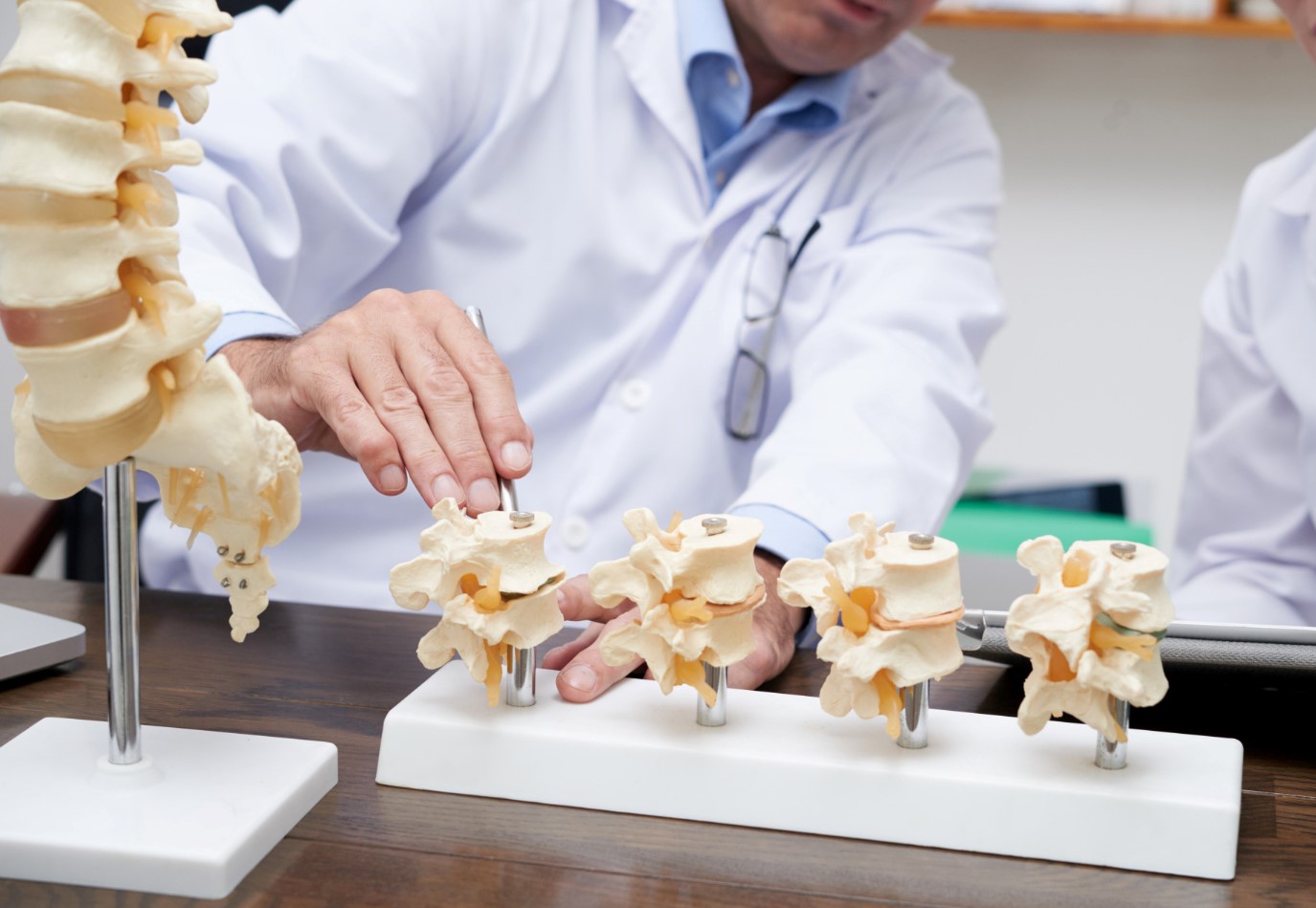
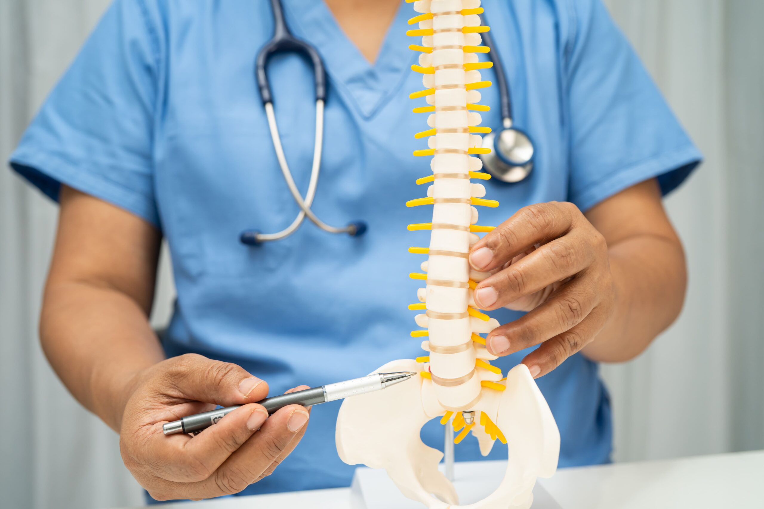
Complications if Left Untreated

Without appropriate treatment, spondylolisthesis may progressively worsen. Understanding potential complications emphasises the importance of proper management.


Continued vertebral slippage can worsen spinal stenosis (narrowing of the spinal canal), further compressing nerves. This progression may lead to chronic pain that becomes increasingly difficult to manage with conservative measures.


Prolonged nerve compression may cause permanent nerve damage, resulting in persistent numbness, weakness, or pain in the legs. Once nerve damage becomes established, full recovery becomes less likely even with treatment.


Quality of life may deteriorate as mobility decreases. Patients may find themselves unable to participate in activities they previously enjoyed. Work capacity may diminish, potentially affecting career and financial stability.


In cases with significant slippage, spinal deformity (abnormal curvature or alignment of the spine) may develop, affecting posture and overall spinal balance and leading to compensatory changes in other spinal segments.

Outcomes differ amongst patients based on individual health factors.

An orthopaedic surgeon or neurosurgeon can assess your specific situation and recommend a suitable treatment approach for you.

Prevention

Whilst not all cases of spondylolisthesis can be prevented, certain strategies may reduce risk and slow progression in those already affected.

Maintaining Core Strength

Strong abdominal and back muscles (the muscles in your stomach area and along your spine) support the spine and reduce stress on vertebrae. Regular exercise focusing on core stability can provide ongoing protection.

Proper Lifting Techniques

Lift with your legs rather than your back. Keep objects close to your body. Avoid twisting whilst lifting. These habits protect the spine during daily activities and occupational tasks.

Weight Management

Maintaining a healthy body weight reduces mechanical stress on the lumbar spine (the lower part of your back). Weight loss can provide symptomatic relief and slow degenerative changes.

Activity Modification for Athletes

Young athletes participating in high-risk sports (such as gymnastics, football, or weightlifting) should follow proper training techniques and avoid overtraining. Adequate rest periods allow the spine to recover from repetitive stress.

Ergonomic Awareness

Proper workstation setup and regular movement breaks may be beneficial for those with sedentary occupations. Standing desks and ergonomic chairs can help reduce spinal strain.

Frequently Asked Questions

Is spondylolisthesis the same as a slipped disc?

No, these are different conditions. Spondylolisthesis involves one vertebra slipping forward over another vertebra. A slipped disc (herniated disc) occurs when the soft cushioning material between your vertebrae pushes out of place. Whilst both conditions can cause back pain and leg symptoms, they involve different structures and may require different treatment approaches. Your orthopaedic surgeon or neurosurgeon can distinguish between these conditions through examination and imaging.

Can spondylolisthesis heal on its own?

Outcomes differ amongst patients based on individual health factors. In some cases, particularly in young patients with acute pars defects (spondylolysis—a stress fracture in part of the vertebra), healing is possible with appropriate rest and bracing. The bone may heal over several months with proper treatment. Once vertebral slippage has occurred, the vertebra does not typically return to its original position without surgical intervention. Conservative treatment focuses on managing symptoms and preventing progression rather than reversing the slippage.

Will I need surgery for spondylolisthesis?

Some patients with spondylolisthesis manage their condition without surgery. Conservative treatments, including physical therapy, medications, and lifestyle modifications, can provide relief for some patients. Your orthopaedic surgeon or neurosurgeon may consider surgery when:

  • Conservative measures do not provide sufficient relief after an adequate trial
  • Neurological symptoms progress
  • Significant instability exists

Your orthopaedic surgeon or neurosurgeon can discuss options based on your individual circumstances and help you make an informed decision.

How long is the recovery after spondylolisthesis surgery?

Response times vary depending on your specific condition. Recovery time varies depending on the specific procedure performed and individual factors. Minimally invasive procedures (where the surgeon makes smaller incisions and uses instruments) can allow faster recovery than traditional open surgery. Some patients begin walking within a day or two of surgery. Return to desk work can occur within four to six weeks. Full recovery, including return to strenuous activities, may take three to six months. Your surgical team can provide guidance throughout your recovery.

Can I still exercise with spondylolisthesis?

Yes, exercise can be beneficial for spondylolisthesis patients. Low-impact activities such as swimming, walking, and stationary cycling are usually well-tolerated. Core strengthening exercises can play a valuable role in supporting spinal health. Activities involving repetitive hyperextension (bending backwards repeatedly, as in gymnastics or certain weightlifting movements) should be avoided or modified. Your physiotherapist can design a safe exercise programme tailored to your condition.

What happens during a consultation with an orthopaedic surgeon or neurosurgeon for spondylolisthesis?

During your consultation, the orthopaedic surgeon or neurosurgeon will:

  • Review your medical history and discuss your symptoms in detail
  • Conduct a physical examination to assess your spine, nerve function, and overall condition
  • Review previous imaging studies (such as X-rays or MRI scans) and order additional tests if needed
  • Explain your diagnosis and discuss treatment options
  • Answer your questions and develop a treatment plan suited to your specific needs
Is spondylolisthesis hereditary?

Genetic factors do play a role in some types of spondylolisthesis. Certain inherited characteristics affecting bone structure and spinal anatomy may predispose individuals to this condition. Spondylolisthesis may occur among family members of affected individuals. If you have close relatives with spondylolisthesis, informing your doctor is helpful, particularly if you develop back symptoms.

How is the severity of spondylolisthesis measured?

Spondylolisthesis is graded using the Meyerding classification system, which measures the percentage of vertebral slippage. This grading helps guide treatment decisions, though symptom severity doesn’t always correlate directly with the degree of slippage. Your doctor can recommend treatment based on your specific symptoms, activity level, and overall health profile.

Conclusion

Spondylolisthesis is a spinal condition ranging from asymptomatic cases discovered incidentally to presentations requiring surgical intervention. Understanding your diagnosis can empower you to participate actively in treatment decisions.


Treatment approaches offer options for patients based on their individual conditions. Some individuals can achieve symptom relief with conservative measures, including physical therapy, pain management, and lifestyle modifications. When surgery becomes necessary, techniques can provide outcomes with faster recovery times.


Successful management involves accurate diagnosis, appropriate treatment selection, and patient commitment to rehabilitation. With proper care, some patients with spondylolisthesis can continue leading active, fulfilling lives.

Take the First Step Towards Better Health

Living with spondylolisthesis can be challenging, but you don’t have to face it alone. Talk to our Orthopaedic Surgeon or Neurosurgeon, who can diagnose and treat spondylolisthesis using evidence-based approaches.

Dr Ng Zhi Xu

Dr Ng Zhi Xu

Senior Consultant Neurosurgeon

Dr Ng is a fully registered specialist in Neurosurgery, with sub-specialty interests in neurotrauma, neuro-oncology, and spine surgery.

  • Bachelor of Medicine and Bachelor of Surgery (MBBS) from Yong Loo Lin School of Medicine, National University of Singapore 2007
  • Basic Surgical Training at TTSH and KTPH 2008-2010
  • Basic Neurosurgical training NUH and KTPH 2010-2012 (Service Registrar)
  • Advanced Neurosurgical training and residency NUH 2013 – 2017
  • Fellowship training in Neurosurgery at Addenbrooke’s Hospital, Cambridge, United Kingdom from 2015 – 2016
  • Intercollegiate Fellowship of Royal Colleges of Surgery (FRCSGlasg) for Neurosurgery, 2016

During his term in KTPH, he developed a deep interest in teaching and organized numerous General Practice (GP) and nursing forums to promote the sharing of common neurosurgical conditions. While there, he was also the director for residency teaching in KTPH.

Dr Ng has done research collaborations with both National Neuroscience Institute (NNI) and NUH. He currently has ongoing research grants with NUH, which focuses on developing modified stem cell treatment for resistant brain cancers.

Dr Roy Koh Kiok Miang

Dr Roy Koh Kiok Miang

Senior Consultant Neurosurgeon

Dr Roy Koh has expertise to manage a wide variety of neurosurgical and spinal problems. He was the first Neurosurgical Spine Consultant at KTPH and also performed Singapore’s first minimally invasive removal of a Spinal Tumor.

  • MBBS, National University of Singapore (NUS) in 2000
  • Basic Surgical Training in 2004
  • MRCS (Edin) and Master in Medicine(Surgery) in 2006
  • Neurosurgical Advanced Surgical training in 2008
  • Fellowship with Neurosurgical Department in Addenbrookes Hospital, Cambridge

Dr Koh has been active in pursuing his love for neurosurgery and spine surgery. He has also been an invited surgeon to places like India and visiting specialist surgeon for the Vietnamese Neurosurgical Congress in 2013, showcasing his new minimally invasive spine techniques to our neighbours.

Dr Koh is an Adjunct Assistant Professor in the Yong Loo Lin School of Medicine, where he is an MBBS examiner, and actively involved in training of the next generation of doctors.

Dr Victor Wang Tzong-Jing

Dr Victor Wang Tzong-Jing

Senior Consultant Orthopaedic Surgeon, Spine & Orthopaedic Surgery

Dr Victor Wang is a fellowship-trained Consultant Orthopaedic Surgeon who sub-specialises in Spine surgery.

  • MBBS (Singapore)
  • MRCS (Edin)
  • MMed (Ortho)
  • FRCSEd (Ortho)

His practice interests include Minimally invasive spine surgery, Navigation/Robot-assisted spine surgeries, Intervertebral disc-related diseases and Adult Spinal Deformities.

A keen believer in the application of technology in evidencebased practice, Dr Wang has vast experience in utilising Computer Navigation for major spine surgeries.

Our clinics are under the following

insurance panels

For Singaporeans, Singapore Permanent Residents and Foreigners.
Please speak to our friendly clinic staff about using your insurance plans.

Reach Out To Us

We are committed to providing a one-stop experience, where you’ll receive a meticulous consultation and high-quality service in a comfortable environment.

Get Started

Book An Appointment


    image

    For faster response, Call Us

    (65) 6733 3383