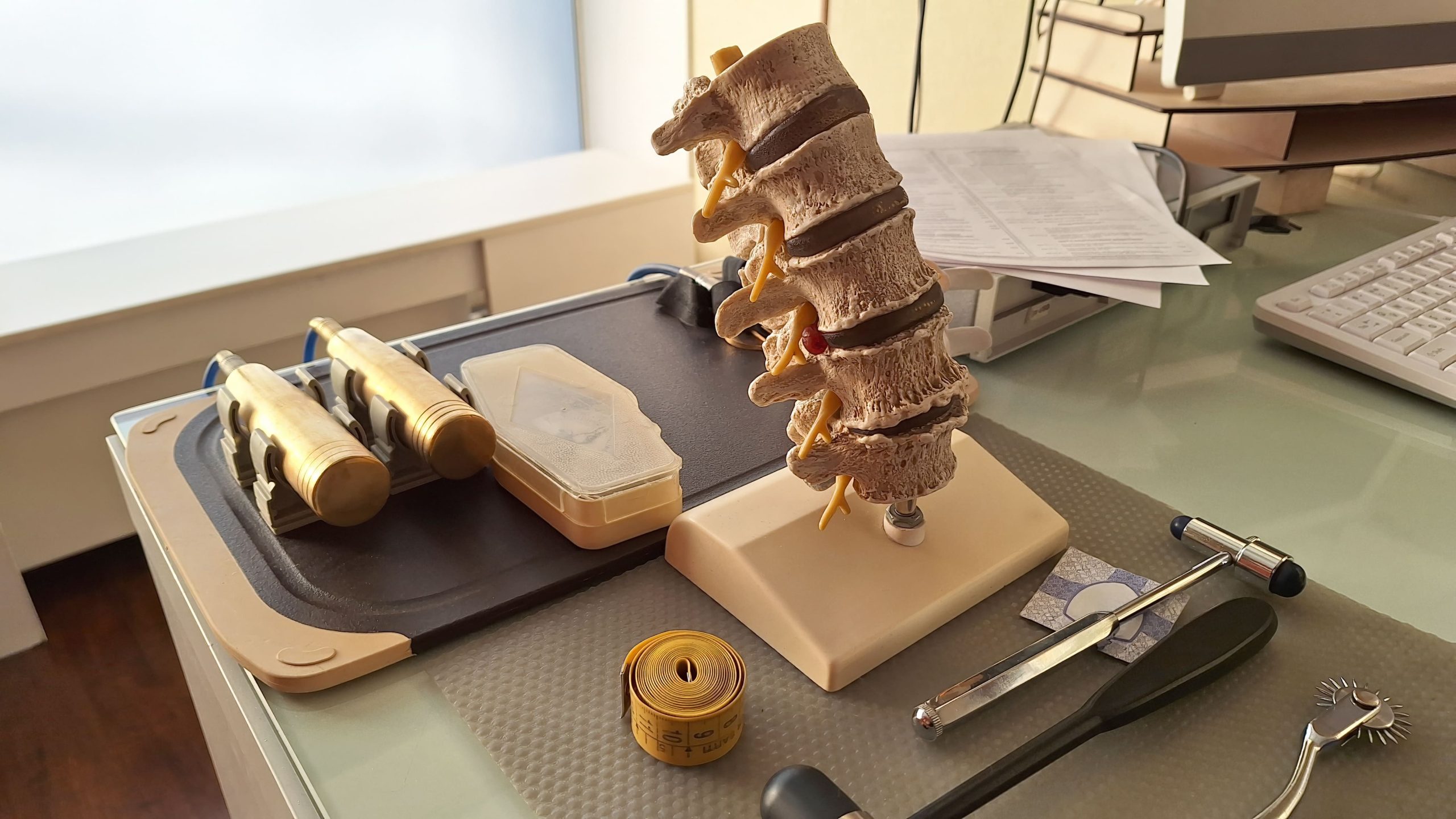
Chronic back pain that worsens with standing but improves when sitting or lying down is a hallmark sign of adult spinal deformity, a condition that disrupts the spine’s natural alignment, posture, and balance. Stemming from either progressed adolescent conditions or age-related spinal degeneration, this disorder encompasses distinct structural patterns such as scoliosis, kyphosis, and lordosis.
Over time, these structural changes create unique mechanical stresses on the body, often forcing adults to unconsciously alter their posture and gait, which can lead to secondary joint issues.
Types of Adult Spinal Deformity
Adult spinal deformity manifests in several distinct structural patterns, each uniquely impacting alignment, stability, and daily physical function.
- Degenerative Scoliosis: Developing later in life from asymmetric disc degeneration and arthritis, this condition causes a gradual sideways curvature that most commonly impacts the lumbar spine.
- Adult Idiopathic Scoliosis: This pattern represents adolescent scoliosis that persists into adulthood, where curves may remain stable for decades or progress further due to age-related disc degeneration, facet joint deterioration, and decreased bone density.
- Kyphosis in Adults: Characterised by an excessive forward rounding of the upper back, severe kyphosis stems from factors like vertebral fractures or disc disease and can compromise balance and, in more severe cases, reduce lung capacity.
- Sagittal Imbalance: This highly disabling condition occurs when the spine loses its front-to-back alignment, shifting the head and trunk forward and forcing constant, exhausting muscular effort to remain upright.
Recognising Symptoms of Adult Spinal Deformity
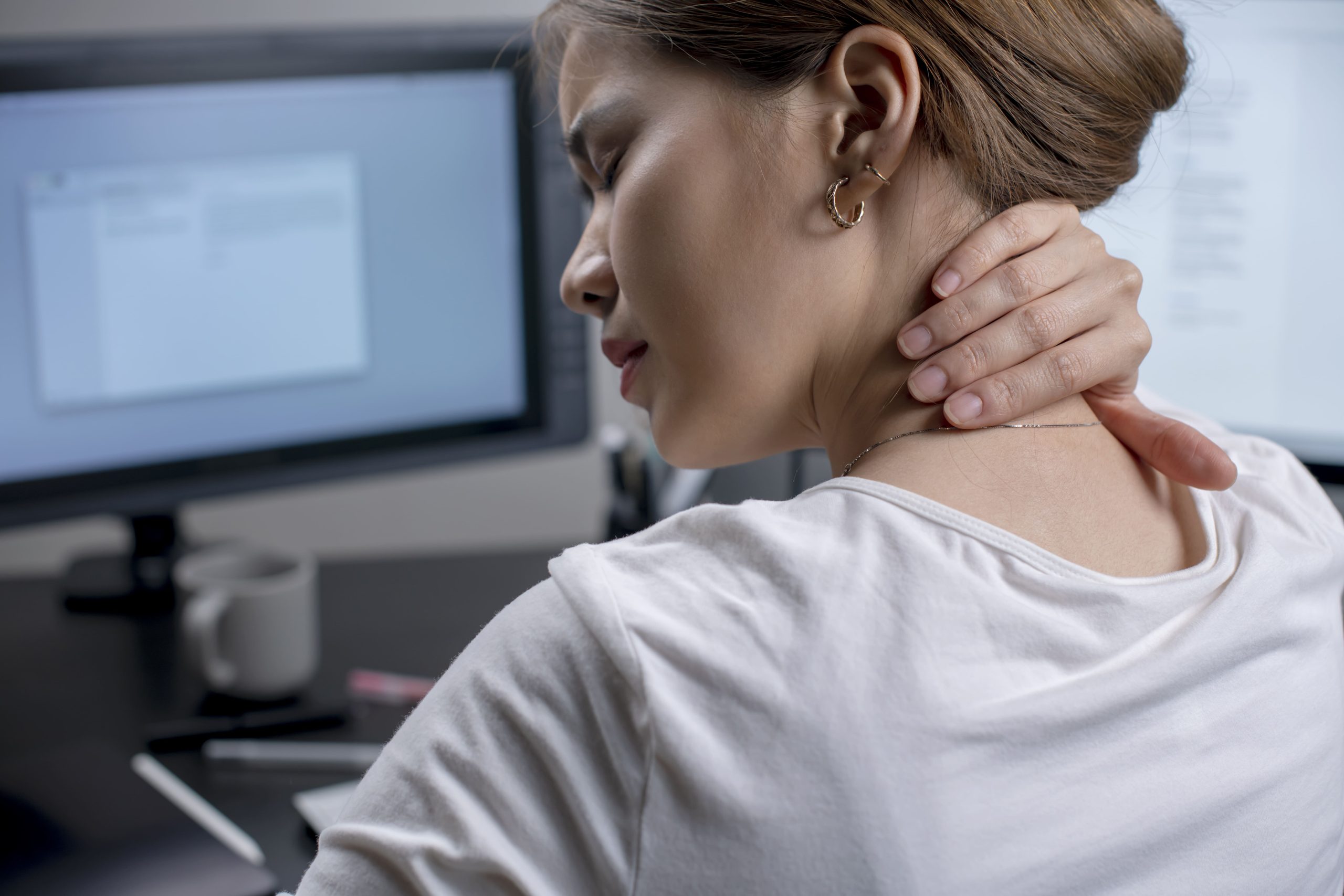
Adult spinal deformity manifests through a combination of mechanical pain, nerve compression, and visible structural changes that impact daily mobility.
- Variable Back Pain: Often localised or radiating into the legs, this mechanical pain characteristically worsens during standing or walking and improves when sitting or lying down.
- Nerve-Related Leg Symptoms: Spinal compression can cause sharp, shooting nerve pain (radiculopathy) or leg heaviness and cramping while walking (neurogenic claudication) that is relieved by leaning forward, sitting, or resting.
- Visible Postural Changes: As alignment shifts, patients may notice uneven shoulders, an asymmetric waistline, a protruding ribcage, or a forward-leaning posture that requires bending the knees to maintain balance.
- Progressive Functional Decline: The structural shift frequently leads to reduced walking tolerance, difficulty climbing stairs, trouble looking straight ahead, and an inability to lie flat comfortably.
What Causes Adult Spinal Deformity
Adult spinal deformity is primarily driven by age-related structural breakdown, bone weakness, or secondary mechanical changes that compromise the spine’s stability.
Degenerative Changes
As spinal discs dry out and lose height over time, they often wear down unevenly, causing adjacent vertebrae to tilt toward the collapsed side. This structural shift is compounded by facet joint arthritis, which creates uneven joint surfaces that encourage abnormal curvature across multiple spinal levels.
Osteoporosis and Vertebral Fractures
Weakened bone density can lead to compression fractures that turn standard vertebrae into wedge-shaped structures, fundamentally altering spinal alignment. While an isolated fracture might cause minimal change, multiple fractures frequently result in severe forward rounding. Postmenopausal women face a particularly elevated risk due to accelerated bone loss, though older men are also significantly affected.
Previous Spinal Surgery
Surgical procedures that fuse spinal segments redirect mechanical stress to the remaining mobile joints above and below the treated area. Over time, this transferred load can accelerate adjacent segment breakdown in some patients or alter the spine’s natural mechanics, sometimes resulting in conditions like flatback syndrome.
Neuromuscular Conditions
Disorders affecting neuromuscular control, such as Parkinson’s disease, muscular dystrophy, and post-polio syndrome, can disrupt the muscular and neurological forces that maintain spinal alignment. In Parkinson’s disease specifically, a combination of muscular rigidity, dystonia, and impaired proprioception contributes to progressive postural deformity.
Diagnostic Evaluation Process
The diagnostic evaluation relies on a combination of physical assessments and advanced imaging to fully map the spine’s structural alignment and identify underlying nerve or bone issues.
- Clinical Examination: Specialists evaluate standing posture, gait, and forward bending to check for rotational curves, while using a structural plumb line to quantify front-to-back balance.
- Standing Full-Spine X-rays: As the cornerstone of imaging, these long-cassette films capture the entire spine to calculate the Cobb angle for curve severity and assess global sagittal balance.
- MRI Scanning: This detailed imaging provides clear views of soft tissues, allowing specialists to pinpoint nerve compression, disc herniations, and spinal canal narrowing.
- CT and CT Myelography: These scans offer intricate views of bone architecture for surgical planning, with myelography using contrast fluid to visualise nerves if an MRI is not an option.
- Bone Density Testing: This test screens for underlying osteoporosis, as poor bone quality directly influences the choice of surgical hardware and may require pre-treatment bone-strengthening medication.
Non-Surgical Treatment Approaches
Conservative, non-surgical therapies focus on relieving chronic pain, improving daily mobility, and supporting the spine’s altered mechanics without invasive intervention.
Physical Therapy
Targeted exercise programmes provide dynamic stability to a structurally compromised spine by focusing on core strengthening, localised flexibility, and tailored posture training. For patients with specific imbalances, interventions like hip flexor stretching, thoracic extension exercises, or low-impact aquatic therapy help reduce spinal loading and prevent compensatory joint tightness.
Pain Management
Medical management utilises a combination of tailored oral medications — ranging from anti-inflammatories to nerve-specific drugs — and targeted localised injections to disrupt chronic pain cycles. Minimally invasive procedures such as epidural steroid or facet joint injections can help reduce inflammation and provide pain relief, which may allow patients to more comfortably participate in physical rehabilitation. The duration and extent of relief vary between individuals.
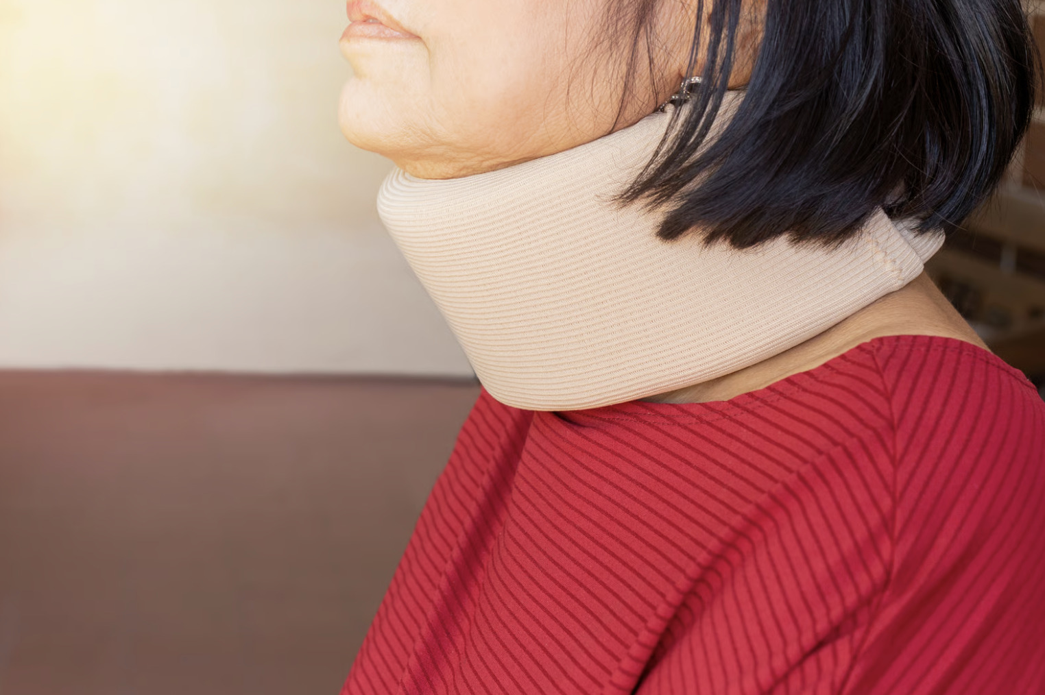
Bracing
While external spinal orthoses cannot permanently restructure or straighten a mature adult spine, they provide valuable external support that reduces the muscular workload required to stand upright. Custom-moulded thoracolumbar braces distribute physical forces across a broad area, and may help reduce mechanical fatigue and discomfort during daily activities, particularly in the short to medium term.
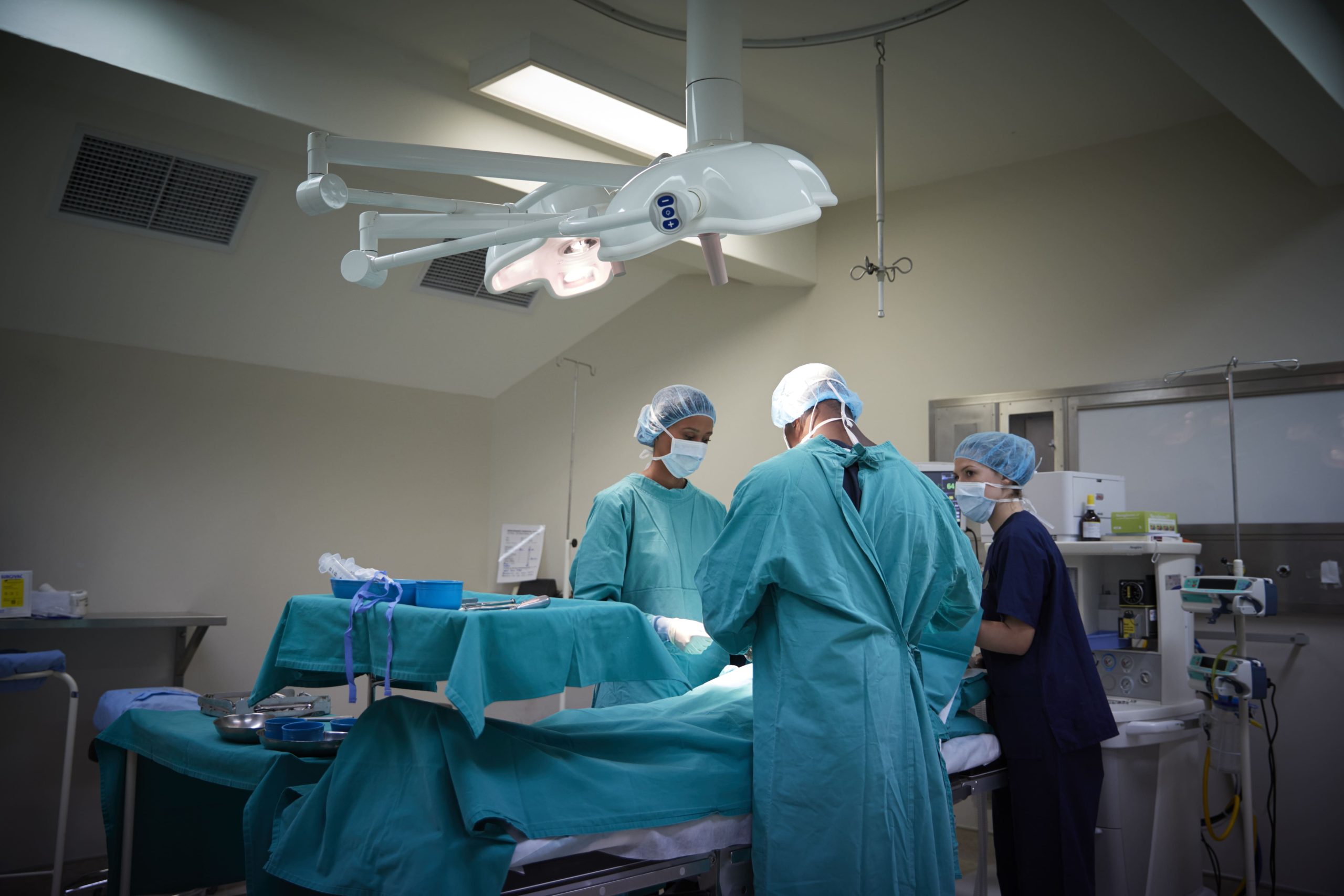
Surgical Treatment Options
Surgical intervention addresses deep structural imbalances that fail to respond to conservative care, aiming to restore proper alignment, relieve nerve pressure, and stabilise the spine.
Decompression Surgery
When compressed nerves cause debilitating leg symptoms without causing global spinal instability, targeted procedures like a laminectomy or foraminotomy are utilised to remove bone or enlarge nerve pathways. These focused interventions successfully relieve direct nerve pressure and associated pain, though they do not correct the underlying structural curvature of the spine.
Spinal Fusion
This procedure joins affected vertebrae together using bone graft material and rigid instrumentation — such as rods and screws — to permanently eliminate painful motion at unstable segments. Surgeons can approach this reconstruction from the back (posterior), front or side (anterior/lateral), or use a combined approach to achieve significant alignment correction and restore mechanical function.
Osteotomy Procedures
For rigid, unyielding spinal curves, surgeons must strategically remove precise sections of bone to reshape and realign the spine into a more natural position. Depending on the severity of the deformity, these techniques range from removing posterior elements and facet joints (posterior column osteotomy) to removing the pedicles and a wedge of vertebral bone (pedicle subtraction osteotomy), up to complete resection of an entire vertebral body (vertebral column resection) for the most severe deformities.
Minimally Invasive Techniques
Modern approaches, such as lateral interbody fusions, utilise advanced tubular retractors to reach the disc space through the side of the body, significantly reducing deep muscle damage and blood loss. While robotic-assisted navigation further improves hardware placement accuracy and speeds up early recovery, highly complex or extensive spinal deformities may still require traditional open surgery.
Recovery and Rehabilitation After Surgery
Post-operative recovery involves a phased transition from safe hospital mobilisation to long-term bone healing and physical rehabilitation.
- In-Hospital Mobilisation: A typical hospital stay for adult spinal deformity surgery ranges from 3 to 7 days, depending on the complexity of the procedure, with early movement encouraged within the first 24 to 48 hours under physical therapy guidance alongside multi-modal pain management.
- Initial Home Healing: The first few weeks at home prioritise wound care, wearing a protective brace, and a gradual increase in walking while strictly avoiding bending, twisting, and lifting.
- Long-Term Bone Fusion: Achieving a solid bone fusion typically takes between 6 and 12 months or longer, requiring proper nutrition, strict compliance with movement restrictions, and the avoidance of smoking and nicotine.
- Structured Rehabilitation: Once early healing is confirmed, formal physical therapy begins to gradually rebuild core strength and endurance, though achieving the full benefits of surgery can take a year or more.
Managing Daily Life with Spinal Deformity
Activity Modification
Understanding which positions and activities aggravate symptoms allows strategic planning. Breaking prolonged standing into shorter intervals, using supportive seating, and maintaining good sleep posture all contribute to symptom management. Assistive devices — walking sticks, rollators, reaching tools — maintain independence while reducing spinal stress.
Exercise Selection
Low-impact activities preserve fitness without excessive spinal loading. Swimming and water aerobics provide cardiovascular conditioning. Stationary cycling in an upright position suits many patients. Walking programmes, adjusted for individual tolerance, maintain functional capacity.
Workplace Adaptations
Ergonomic assessment identifies modifications that reduce occupational strain. Adjustable desks allowing position changes, supportive seating, and strategic break scheduling help those who must work with spinal deformity. Some occupations prove incompatible with significant deformity, necessitating role changes.
When to Seek Professional Help
- Progressive difficulty with walking or standing upright
- New or worsening leg weakness or numbness — and especially any bowel or bladder dysfunction (such as difficulty urinating, incontinence, or altered sensation in the groin area) — require urgent or emergency medical evaluation and should not be delayed
- Pain that disrupts sleep or daily activities despite basic measures
- Visible postural changes worsening over time
- Inability to look straight ahead while walking
- Pain requiring increasing medication for control
- Difficulty performing work or daily tasks
Commonly Asked Questions
Does adult spinal deformity always get worse over time?
Progression varies considerably. Degenerative curves typically worsen gradually, while some adult idiopathic curves remain stable for years. Factors influencing progression include curve location and magnitude, bone density, and overall spinal health. Regular monitoring through clinical examination and periodic imaging tracks curve behaviour.
Can exercise straighten a curved spine in adults?
Exercise cannot reverse the underlying bony structural spinal curvature in adults — the vertebrae and discs will not undergo structural remodelling through exercise alone. However, scoliosis-specific exercises may help reduce the postural component of curvature and slow progression. Exercise remains highly beneficial for reducing pain, improving function, and strengthening supporting muscles. The goals differ from adolescent treatment, where bracing and exercise may influence a growing spine.
How do I know if I need surgery for spinal deformity?
Surgery becomes appropriate when non-surgical measures fail to adequately control symptoms, when neurological function deteriorates, or when deformity progression threatens future function. The decision involves weighing symptom severity, functional limitations, overall health, and surgical risks. Most patients with adult spinal deformity manage successfully without surgery.
What is the recovery time after spinal deformity surgery?
Recovery timelines depend on surgical extent. Limited decompression procedures allow return to normal activities within weeks. Major reconstructive surgery with fusion requires months for bone healing and up to a year for full functional recovery. Individual factors including age, fitness, and procedure complexity influence these timelines.
Will spinal deformity affect my ability to work?
Impact on work capacity depends on deformity severity, symptom control, and occupational demands. Many people with spinal deformity continue working with appropriate modifications. Physically demanding occupations may require role adjustments. Severe deformity with poor symptom control can limit work capacity significantly.
Next Steps
Accurate diagnosis establishes the deformity pattern and identifies contributing factors essential to guiding care. Exercise, injections, and surgery each address different aspects of the condition — treatment intensity should match the clinical picture.
For progressive symptoms or neurological changes such as leg weakness or numbness, prompt surgical evaluation is warranted. New or worsening bowel or bladder dysfunction requires emergency medical evaluation and should not be delayed, as it may indicate cauda equina syndrome — a surgical emergency.
If you are experiencing back pain that worsens with standing, difficulty maintaining an upright posture, or leg symptoms such as weakness, numbness, or claudication, consult an orthopaedic surgeon for a comprehensive spinal evaluation and a discussion of appropriate treatment options.