Skull Base Tumour Excision

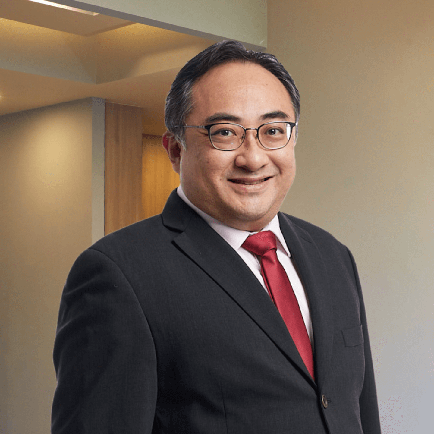
Dr Roy Koh Kiok Miang
Consultant Neurosurgeon

What Is Skull Base Tumour Excision?

Skull base tumour excision is a complex surgical procedure aimed at removing tumours at the skull’s base. This region is a critical area where the bottom part of the skull meets the top part of the spinal column, and it encompasses various nerves and blood vessels that serve the brain, head, neck, and other parts of the body.

The intricacy of this area means that tumours within this region pose significant challenges for treatment. Skull base tumour excision aims to remove the tumour while minimising damage to surrounding tissues, nerves, and blood vessels.

Types of Skull Base Tumours

These are some of the common types of skull base tumours:

Chordomas

Chordomas are rare, slow-growing tumours that typically occur at the base of the skull or along the spine. They arise from notochord remnants, a structure present during embryonic development.

Meningiomas

Meningiomas are the most common type of primary brain tumours arising from the meninges, the membranes that cover the brain and spinal cord. Most meningiomas are benign and grow slowly.

Pituitary Adenomas

These are benign tumours that develop in the pituitary gland, a small gland at the base of the brain that plays a major role in regulating vital body functions and hormones. They can affect hormone levels and cause a variety of symptoms.

Schwannomas

Also known as acoustic neuromas, these are benign tumours that develop from the Schwann cells surrounding nerves. The most common type affects the vestibular nerve, leading to hearing loss and balance issues.

Craniopharyngiomas

Craniopharyngiomas are benign tumours that develop near the pituitary gland and can affect hormone production, leading to various symptoms. They are commonly diagnosed in children and young adults.

Symptoms

Symptoms vary widely and depend on the tumour’s location, size, and the affected structures. These are some of the most common symptoms:

  • Headaches, often worsened by lying down or early in the morning
  • Changes in vision, such as blurred or double vision
  • Loss of sensation or altered sensation in the face
  • Weakness or paralysis on one side of the face
  • Hearing loss or ringing in the ears
  • Difficulty swallowing

In cases where the tumour affects the pituitary gland:

  • Changes in the menstrual cycle
  • Unexpected lactation
  • Changes in libido
  • Unexplained weight gain or loss
  • Fatigue
  • Mood swings

  • Persistent nasal congestion or nosebleeds, especially in tumours near the nasal passages
  • Difficulty with balance or coordination
  • Personality changes

Preparing for Skull Base Tumour Excision

Preparation for skull base tumour excision involves several key processes to ensure the safety and success of the surgery.

Detailed Medical Evaluation

  • Comprehensive medical history review and physical examination.
  • Assessment of the patient’s overall health to ensure they are fit for surgery.

Imaging Studies

Magnetic resonance imaging (MRI) and computed tomography (CT) scans determine the tumour’s exact location, size, and extent, as well as its relationship to surrounding structures.

Neurological Testing

Tests to assess neurological function, including hearing tests and vision tests, depending on the tumour’s location.

Endocrine Evaluation

If the tumour is near the pituitary gland, tests to assess hormone levels may be conducted.

Patient Counselling

  • Patients are informed about the details of the procedure, potential risks and complications, the recovery process, and the expected outcomes.
  • Discussion about the need for postoperative care, including rehabilitation and follow-up visits.

Preoperative Instructions

  • Guidance on medications, fasting, and other preparations before the surgery.
  • Arrangements for postoperative care and support.

Surgical Techniques

The surgical removal of skull base tumours is highly complex, and techniques have evolved significantly, with a focus on minimising trauma to surrounding tissues and improving patient outcomes.

These are some of the primary surgical techniques used in skull base tumour excision:

Open Surgery

Traditional open surgery involves making a large incision to access the tumour. This may require removing part of the skull or facial bones for direct access.

Minimally Invasive Endoscopic Surgery

This technique uses an endoscope, a thin, flexible tube with a light and camera at the end, to access and remove the tumour through small incisions or natural openings, such as the nostrils.

Microsurgical Techniques

Microsurgery involves using a microscope to enhance the surgeon’s view of the small and complex structures at the skull base. This allows for precise tumour removal with less damage to surrounding tissues.

Stereotactic Radiosurgery

Stereotactic radiosurgery uses focused radiation beams to target and destroy the tumour cells without an incision. It is often used for tumours that are difficult to reach surgically or for patients who cannot undergo traditional surgery.

Risks and Complications of Surgery

Like all surgical procedures, skull base tumour excision carries some risks and potential complications.

  • Infection: As with any surgery, there is a risk of infection at the incision site or within the skull. Infections are treated with antibiotics but can extend hospital stays and recovery.
  • Bleeding: There is a risk of significant bleeding during or after surgery, given the area’s rich blood supply. In rare cases, a blood transfusion or additional surgery may be needed to control bleeding.
  • Neurological Damage: Given the proximity to vital nerves and brain structures, there is a risk of damage leading to loss of smell, vision impairments, facial paralysis, or difficulties with balance and coordination.
  • Cerebrospinal Fluid (CSF) Leak: Surgery may result in a leak of CSF, the fluid surrounding and protecting the brain and spinal cord. This can lead to meningitis, a severe infection of the brain’s protective membranes, and may require additional surgery to repair.
  • Hormonal Imbalances: If the tumour is near or involves the pituitary gland, there may be changes in hormone levels requiring long-term medication to manage.
  • Recurrence: There is always a risk that some tumour cells may remain, leading to the tumour’s recurrence. Regular follow-up is necessary to monitor for any signs of recurrence.

Dr Roy Koh Kiok Miang

Consultant Neurosurgeon

Dr Roy Koh has expertise to manage a wide variety of neurosurgical and spinal problems. He was the first Neurosurgical Spine Consultant at KTPH and also performed Singapore’s first minimally invasive removal of a Spinal Tumor.

  • MBBS, National University of Singapore (NUS) in 2000
  • Basic Surgical Training in 2004
  • MRCS (Edin) and Master in Medicine(Surgery) in 2006
  • Neurosurgical Advanced Surgical training in 2008
  • Fellowship with Neurosurgical Department in Addenbrookes Hospital, Cambridge

Dr Koh has been active in pursuing his love for neurosurgery and spine surgery. He has also been an invited surgeon to places like India and visiting specialist surgeon for the Vietnamese Neurosurgical Congress in 2013, showcasing his new minimally invasive spine techniques to our neighbours.

Dr Koh is an Adjunct Assistant Professor in the Yong Loo Lin School of Medicine, where he is an MBBS examiner, and actively involved in training of the next generation of doctors.

Our clinics are under the following

insurance panels

For Singaporeans, Singapore Permanent Residents and Foreigners.
Please speak to our friendly clinic staff about using your insurance plans.

Reach Out To Us

We are committed to providing a one-stop experience, where you’ll receive a meticulous consultation and high-quality service in a comfortable environment.

Get Started

Book An Appointment


    image

    For faster response, Call Us

    (65) 6733 3383

    Skull Base Tumour Excision in Singapore

    Frequently Asked
    Questions

    Can skull base tumours be benign, or are they always cancerous?

    Skull base tumours can be benign (non-cancerous) or malignant (cancerous). While not spreading to other parts of the body, benign tumours can still cause significant symptoms and complications due to their location.

    How long does recovery from skull base tumour excision take?

    Recovery time varies depending on the type of surgery, the size and location of the tumour, and the patient’s overall health. It can range from a few weeks for less invasive procedures to several months for more complex surgeries.

    What are the alternatives to surgery for skull base tumours?

    Alternatives to surgery may include observation for small, asymptomatic tumours, radiation therapy, or stereotactic radiosurgery for tumours that are difficult to remove surgically.