
The patient lies down on an examination table, and the area to be examined is exposed while the rest of the body remains covered. The patient may be asked to raise their arm above their head to allow better access to the breast tissue.
A clear, water-based gel is applied to the breast. This gel helps to eliminate air pockets between the transducer and the skin, which can interfere with the sound waves needed to create the images.
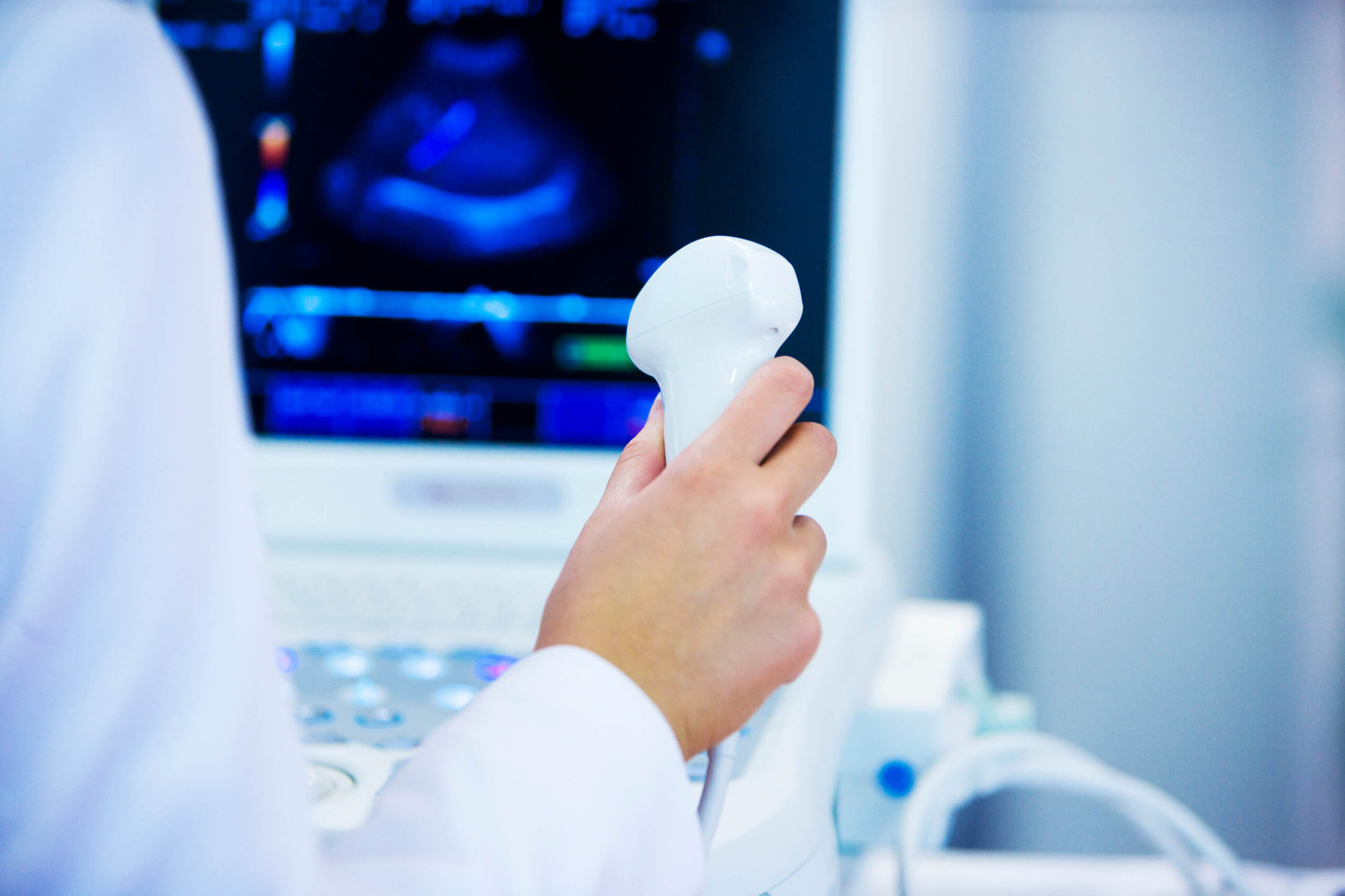
The radiologist or sonographer gently presses the transducer against the skin and moves it over the area of interest. The transducer sends and receives high-frequency sound waves that create live images of the breast tissues on a monitor.
The sonographer might occasionally pause to examine certain images more closely, sometimes capturing specific pictures for later review. Additional images or focus on specific areas may be necessary if any unusual findings are observed.
Once the examination is complete, the gel is wiped off, and the patient can get dressed. There are no post-procedure restrictions, allowing the patient to return to normal activities immediately.
The images from the ultrasound are reviewed by a radiologist, who interprets the data and compiles a report for the patient’s physician. The results are usually available within a few days, and a follow-up appointment may be scheduled to discuss the findings.


