
Lesion Imaging: Accurate imaging studies, such as mammography or ultrasound, locate the lesion and plan the wire placement.
Local Anaesthesia: Local anaesthesia is administered at the insertion site to minimise discomfort.
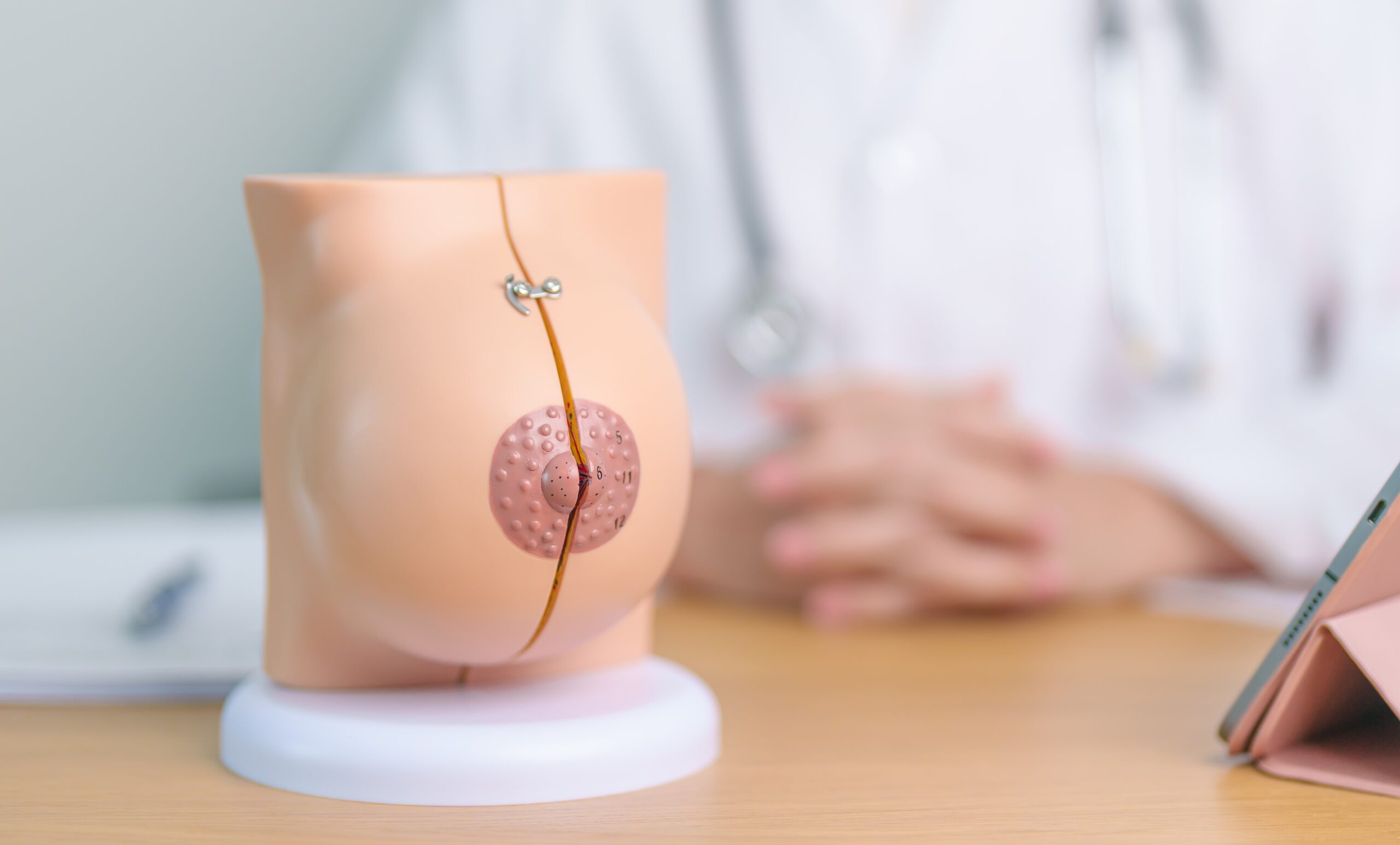
Wire Placement: Under real-time imaging guidance, a radiologist inserts the hookwire through the skin and into or near the lesion. The wire has a hook at the end to anchor it securely within or around the lesion.
Confirmation of Placement: Imaging confirms that the hookwire is correctly positioned relative to the lesion.
Guidance to Lesion: During surgery, the protruding part of the wire guides the surgeon to the lesion.
Lesion Removal: Following the wire, the surgeon excises the lesion, including the hooked end of the wire within the excised tissue, to confirm complete removal.
Assessment of Margins: The excised tissue, along with the hookwire, is sent for pathological examination to assess if the lesion has been fully excised and to evaluate the margins.
Immediate Post-operative Care: Post-operative care includes monitoring for any complications at the wire insertion site and managing pain.
Follow-up: The patient will have follow-up visits to review the pathology report, assess healing, and discuss any further treatment necessary based on the surgical and pathological findings.


